Ultrasound: answers to popular questions
Many hidden health problems can be detected by ultrasound. But in order to get complete undistorted information about the state of internal organs, it is important to properly prepare for the procedure. It is also useful for patients to know about the technology, features and purposes of ultrasound diagnostics. We will try to give short and clear answers to popular questions about ultrasound.
What is an ultrasound scan?
Ultrasound examination (ultrasound) is a non-invasive diagnostic method that allows you to obtain information about the position and condition of internal organs. Visualization of areas of the human body hidden from the eyes is achieved through exposure to ultrasonic waves.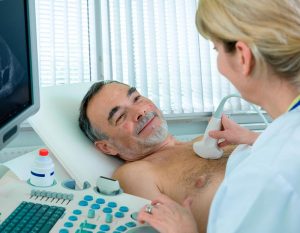
A special transducer emits echoes of a specific frequency and receives the returning reflections. Sound waves are digitized and displayed on a monitor in real time. To study the vessels, a special Doppler ultrasound is performed.
When patients ask themselves what an ultrasound scan is, they want to first of all know how safe this diagnostic method is. During the procedure, radiation hazardous to humans is not used. At high intensity ultrasound cavitation is possible, but this side effect is very rare in medical practice.
How to prepare for an ultrasound scan?
Before an abdominal ultrasound, it is important to reduce the level of gas formation, since the presence of gas bubbles prevents the ultrasound from penetrating to the examined areas. Diagnostics recommend that adult patients follow a special diet for 3 days. It is necessary to avoid the use of foods that can cause fermentation, observe the frequency of meals and the drinking regime.
How to prepare for an ultrasound scan for young patients, pregnant women, people with diabetes? Children will need less time to get ready, since gas is removed from the body faster due to the rapid metabolism. Food intake stops 5-6 hours before the ultrasound scan. Patients with diabetes mellitus and pregnant women can afford a light snack (a few white bread crumbs, tea with honey and lemon, etc.) 2 hours before the procedure.
Before examining the kidneys and bladder, the patient should drink 1.5 liters of fluid one hour before the procedure. If a transabdominal examination of the pelvic organs is planned, then you will need to drink about 0.5 liters of clean water also one hour before visiting the ultrasound office. It is not recommended to chew gum before the procedure, as this increases intestinal motility. It is also worth giving up mouth freshness sprays, cigarettes.
For women, for ultrasound of the pelvis and mammary glands, it is important to choose the right cycle day – usually 5-8 days are recommended for more accurate results.
How is ultrasound done?
Diagnostics using an ultrasound machine is carried out by qualified diagnosticians who know the intricacies of working with the equipment and are able to correctly decipher the information received.
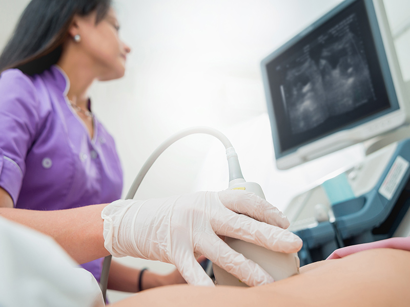
During the procedure, the person must be in a stationary position. A special gel is applied to the skin, which eliminates the formation of an air gap that impedes the transmission and reception of ultrasonic signals. How is ultrasound done for young children who are very restless? Safe sedatives may be used.
During the examination, the diagnostician may ask you to change the position of the body, hold your breath. An ultrasound scan usually takes no more than 20 minutes to diagnose, but in some cases the procedure can be delayed. The results are entered into a special medical protocol.
What does ultrasound show?
The picture obtained during an ultrasound examination most accurately reflects the current state of internal organs and tissues. With the help of ultrasound, you can get information about the size, shape, outlines of organs, identify cysts and other neoplasms, foci of the inflammatory process, uncharacteristic inclusions.
What does vascular ultrasound (dopplerography) show? Such a study allows you to assess the intensity of blood flow, to identify varicose veins, stenosis, blood clots. Ultrasound https://en.wikipedia.org/wiki/Ultrasound of the heart is performed with the aim of confirming or refuting the presence of congenital and acquired defects, changes in large vessels.